


Endodontic treatment focuses on saving natural teeth by treating infections or damage inside the tooth. When left untreated, these issues can lead to more serious complications, including tooth loss. At Elite Dental Associates in Redlands, CA, we use advanced techniques to remove infection, relieve discomfort, and preserve your smile. Schedule a consultation today and let us help restore your oral health!
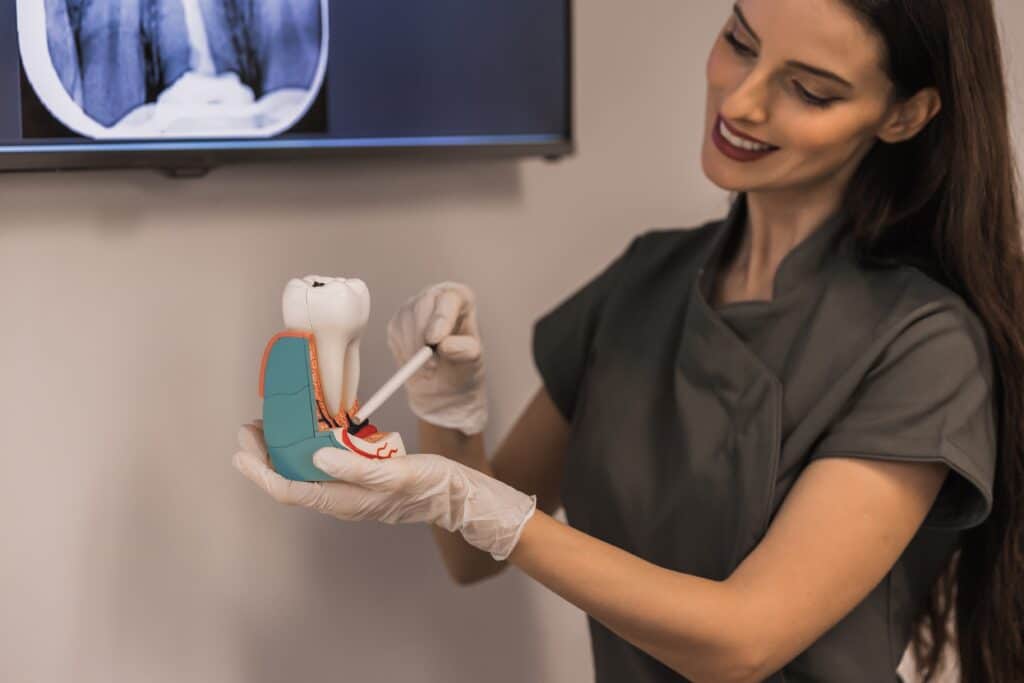
Endodontics is a specialized field of dentistry that focuses on treating issues within the tooth’s pulp and roots. When the inner part of a tooth becomes inflamed or infected, endodontic procedures can help prevent the need for an extraction. These treatments allow patients to keep their natural teeth while eliminating discomfort and improving oral health.
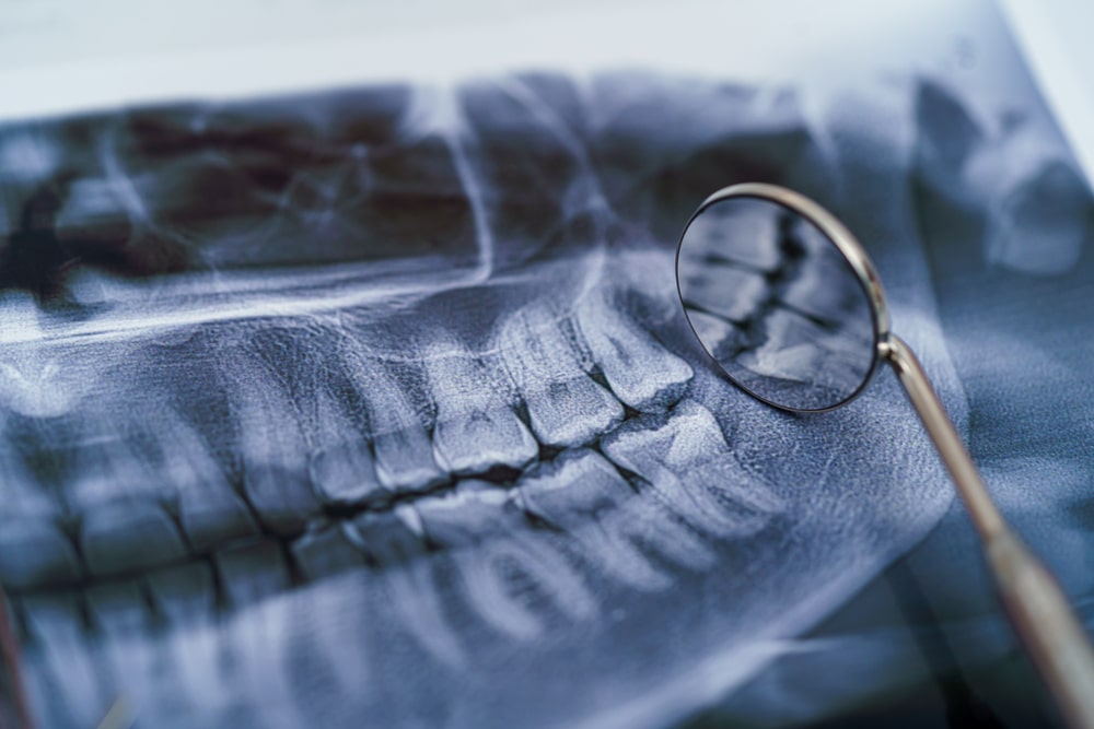
Endodontic treatment is often necessary when an infection reaches the inner layers of a tooth. Early symptoms may be mild, but as the infection spreads, the discomfort can become severe. If you notice any of the following signs, it may be time to see a dentist.
Our team offers a range of endodontic treatments designed to relieve discomfort and restore your natural teeth. Depending on your specific needs, we may recommend one of the following procedures.
A root canal removes infected pulp, cleans the inner canals, and seals the tooth to prevent reinfection. This procedure allows patients to keep their natural teeth while eliminating the source of discomfort. Many people feel relief shortly after treatment and can return to normal activities the next day.
If a previous root canal does not heal properly or becomes reinfected, retreatment may be needed. The procedure involves reopening the tooth, removing old filling material, disinfecting the canals, and resealing the tooth to prevent future infection.
An apicoectomy is a minor surgical procedure used when a root canal alone isn’t enough to eliminate an infection. It involves removing the tip of the tooth’s root along with any infected tissue. This treatment helps preserve the tooth and prevent future problems.
A pulpotomy is commonly performed on children to treat an infection while preserving the healthy part of the pulp. This procedure helps maintain baby teeth until they naturally fall out. It is a great way to prevent discomfort and avoid early tooth loss.
A pulpectomy is needed when the entire pulp inside a tooth is infected. This treatment removes all infected tissue, thoroughly cleans the root canals, and seals the tooth. It is typically followed by a restoration, such as a crown, to protect the tooth.
Endodontic treatment can help save your natural teeth and prevent future dental problems. At Elite Dental Associates in Redlands, CA, we provide expert care to ensure lasting results and a comfortable experience. If you’re dealing with dental discomfort, now is the time to take action. Contact us today to schedule your consultation!